��ʿinsphero GravityTRAP ULA 3D������-3Dϸ������������,insphero3Dϸ������������,3D INSIGHT PLATFORMS��3Dϸ������������--��ʿinsphero
�ͺ�:GravityTRAP
��ϵ��:��ʤ��
��ϵ�绰:18618101725
Ʒ��: ��ʿinsphero
3Dϸ�����������塪����ʿinspheroƷ��
GravityPLUS platform kit�����ţ�CS-06-001�����10��96�װ���
GravityPLUS? and GravityTRAP? Trial Pack�����ţ�CS-00-020�����1��96�װ���
Introductory GravityPLUS and GravityTRAP pack�����ţ�CS-06-010�����3��96�װ壩
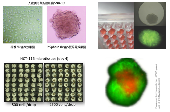
��ʿinsphero��˾������3Dϸ������������wu��ʹ��֧�ܽ���(scaffold-free)��ͨ��������������hanging-drop�� �����ܽ�ϸ����Һ��һ��ϸ�������ϸ�����γ�3D��֯��3D��֯�ʶ�ϸ����״�壬wu������̬ѧ�ϣ������ڹ����Ͼ�����Ȼ��֯���ơ��Ӷ����õ�ģ������ϸ�������������ֱ�۵ķ�ӳϸ������ѧ���ܣ���ȷ�Ĺ�������֯ģ�͡�
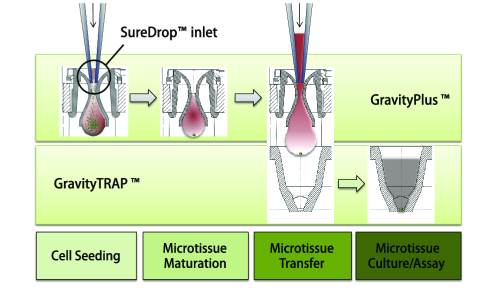
3Dϸ����������Ҫ��GravityPlus���GravityTRAP����ɣ�����ͼ��

GravityPlusTM�壺����ϸ�����η���ר����������ʹϸ���γ�����״��֯��3D״̬������ϸ����Һ��һ��ϸ�������ϸ�������ӵ�GravityPlusTM���У�2-4�죬ϸ����GravityPlusTM���л��γ���֯����״����
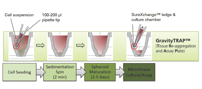
GravityTRAPTM�壺���÷�ճ������������ʹ��֯��wu�����������������������ܣ����ں���ʵ�鼰��⡣����֯�γɺ���ת�Ƶ�GravityTRAPTM���У���ʹ��֯��wu�����������������������ܣ����ں���ʵ�鼰��⡣
Ӧ��ʵ��
3Dϸ�������������Ʒ�Ƽ�Ӧ��
1. 3D�����������γɵ�3D��֯�ʶ�ϸ����״�壬wu������̬ѧ�ϣ������ڹ����Ͼ�����Ȼ��֯���ơ�
2. wu��֧�ܽ��ʣ�ϸ�����������������ܣ��ɶ���֯�����ҩ
3. �����ڱ���ѧ�����ӫ���⣬�ɼ��ATP��LDH��GSH��ϸ��������ָ��
4. 96�װ壬jȷ�ijߴ���ƣ����γɵ���֯������С�����ͨ�������豸���ݣ������ڸ��ں�����(high
content test)
5. �����ڼ����κ�����ϸ����3D��������ϸ��ϵ��ԭ��ϸ������ϸ��������ϸ��������
6. �����ڰ�֢�о���ҩ�������ҩ���л����ѧ(DMPK)�����Բ���(ADME-TOX)��
7. �������ǰ15����ҩ�ͻ���Ʒ��ҵ����ʹ��InSphero��GravityPLUS?ƽ̨��3D��֯����Ʒ��������ISO 9001:2008����
������Ϣ
3D���������婦GravityPLUS platform kit�����ţ�CS-06-001�����10��96�װ壩
3D���������婦GravityPLUS? and GravityTRAP? Trial Pack�����ţ�CS-00-020�����1��96�װ壩
3D���������婦Introductory GravityPLUS and GravityTRAP pack�����ţ�CS-06-010�����3��96�װ壩
InSphero��GravityPLUS��������ϵͳ��GravityTRAP ULA������� |
||||||||||||||||||||||||||||||
|
|
||||||||||||||||||||||||||||||
|
InSphero��2010��ʹ��3D��������������Hanging Drop Technology����������GravityPLUS ��������ϵͳ��InSphero�ڽ������Ƴ�һ����Ʒ��GravityTRAP ULA�塣��ô�������ڽ���3Dϸ������ʵ��ʱ�������ѡ���ģ�����Ͷ�GravityPLUS ��������ϵͳ��GravityTRAP ULA�������¶Աȣ�
��ϸ�ԱȽ������£�
1.��ͬ�㣺 1������96���������壬�������ȸߣ������������й۲⣻ 2�������ڸ�ͨ�����������ں�����������
2.��ͬ�� 1�� GravityPLUS ��������ϵͳ�����������ɣ�GravityPlus �壨���ΰ壩��GravityTRAP�壨�ο�ƽ�װ壩���������ͼ���Ƿֱ���GravityPlus���GravityTRAP������ɣ���ͼʾ��
GravityTRAP ULA��ֻ��һ�����ɣ�GravityTRAP�壨�ο�ƽ�װ壩���������ͼ�����ͬһ���GravityTRAP������ɣ���3Dϸ�����������㣬��ͼʾ��
2��GravityPLUS ��������ϵͳ�����ڼ���allϸ����3Dϸ����������������ϸ����ϸ��ϵ��ԭ��ϸ������ϸ�����Լ���ϸ������������GravityTRAP ULA��ֻ�����������γ��������ϸ��ϵ�ͼ۸�͵�������ϸ��ϵ�� 3��GravityPLUS ��������ϵͳ�۸�ƫ�ߣ���GravityTRAP ULA��۸������ |
3Dϸ������������--��ʿinsphero
3Dϸ������������--��ʿinsphero
3D InSight? Cell-Culture Media
3Dϸ����������������
1. 3Dϸ�������������ר������������Ҫ����ά��3D��֯
2. �����ö�������ά��3D״̬������������֤3D��֯������������ʱ��
3. �����͡�wu���Ϻ�Ԥ��������������ֱ������3D InSight?��֯
������Ϣ
-
����
����
���
CS-07-101
3D InSight? ϸ��ϵά��������
250/500mL
CS-07-100
3D InSight? HepG2��֯ά��������
500mL
CS-07-001
3D InSight? �˸�ϸ��ά��������
250/500mL
CS-07-002
3D InSight? �����ϸ��ά��������
250/500mL
CS-07-005
3D InSight? ���ȵ�ά��������
250/500mL
CS-07-006
3D InSight? �����ȵ�ά��������
250/500mL
Insphero��˾���
InSphero��ͨ��������ʽ3D��֯����(supplier of organotypic, biological
in vitro 3D microtissues)��չ��ҩ���Ե�����Ϳ����̡���ǰ����֪����ǰʮ��ҩ��ͻ���Ʒ�����̲�Ʒ���Զ�ʹ�õ�InSphero��ר����֯������
InSphero 3D Insight?��֯���ڻ�ѧƷӦ�õ�Ч�ܺͶ���������ԣ���������ЧӦ����֢�鵼�Ķ���ЧӦ�������пɹ�ѡ��Ķ�������������֯��Ʒ����������ģ��(toxicology models in the pipeline)��InSphero
3D��֯�ϴ�ͳϸ��ģʽ������Ԥ����������������ʱ��������۸�Ҳ�����ˡ�
InSphero��Jan
Lichtenberg��ʿ, Jens M. Kelm��ʿ��Wolfgang
Moritz��ʿ����2009�����������ʿ��������������ѧԺ(Swiss
Federal Institute of Technology (ETH) Zurich)����������ѧ(University
Zurich)��������˾���ܲ�����Schlieren���������͵¹����з�֧������InSphero��Ʒ�������Һ��ʿ�ѧ����ҵ����������Ʒ��������ISO
9001:2008����
InSphero�з��ŶӾۺ�����֯���̣�ʵ�����Զ���������ѧ��50����ḻ���飬�����ͻ���ҩ�������µġ����߶�Ϥ�������顣
Ӧ�����ף�
Messner S, Fredriksson L, Lauschke VM, Roessger K, Escher C, Bober M, Kelm JM, Ingelman-Sundberg M, Moritz W. (2017) Transcriptomic, Proteomic, and Functional Long-term Characterization of Multicellular Three-Dimensional Human Liver Microtissues. Applied In Vitro Toxicology. doi: 10.1089/aivt.2017.0022.
Proctor WR, Foster AJ, Vogt J, Summers C, Middleton B, Pillings MA, Shienson D, Kijanska M, Stroebel S, Kelm JM, Morgan P, Messner S, Williams D. (2017) Utility of spherical human liver microtissues for prediction of clinical drug-induced liver injury. Arch Toxicol. Aug;91(8):2849-2863. doi: 10.1007/s00204-017-2002-1.
F. Paech, S. Messner, J. Spickermann, M. Wind, A.-H. Schmitt-Hoffmann, A.T. Witschi, B.A. Howell, R.J. Church, J. Woodhead, M. Engelhardt, S. Kr?henb��hl, M. Maurer (2017) Mechanisms of hepatotoxicity associated with the monocyclic ��-lactam antibiotic BAL30072. Arch. Toxicol. doi:10.1007/s00204-017-1994-x..
Hendriks DF, Fredriksson Puigvert L, Messner S, Mortiz W, Ingelman-Sundberg M (2016) Hepatic 3D spheroid models for the detection and study of compounds with cholestatic liability. Sci Rep. 2016 Oct 19;6:35434. doi: 10.1038/srep35434.
Linfeng L., Zhou Q., Voss T.C., Quick K.L., LaBarbera D.V., (2016). High-throughput imaging: Focusing in on drug discovery in 3D. Methods. 2016 Mar 1;96:97-102. doi: 10.1016/j.ymeth.2015.11.013.
Bruderer R., Bernhardt O.M., Gandhi T., Miladinovic S.M., Cheng L.Y., Messner S., Ehrenberger T., Zanotelli V., Butscheid Y., Escher C., Vitek O., Rinner O., and Reiter L. (2015). Extending the limits of quantitative proteome profiling with data-independent acquisition and application to acetaminophen treated 3D liver microtissues. Mol Cell Proteomics. May;14(5):1400-10. doi: 10.1074/mcp.M114.044305.
Kermanizadeh A., Lohr M., Roursqaard M., Messner S., Gunness P., Kelm J.M., Moller P., Ston V., Loft S. (2014). Hepatic toxicology follwing single and multiple exposure of nanomaterials utilising a novel primary human 3D liver microtissue model. Part Fibre Toxicol. Oct 20;11:56. doi: 10.1186/s12989-014-0056-2.
Fruhwurth S., Kovacs W.J., Bittman R., Messner S., Rohrl C., Stangl H. (2014). Differential basolateral-apical distribution of scavenger receptor, class B, type I in cultured cells and the liver. Histochem Cell Biol. Dec;142(6):645-55. doi: 10.1007/s00418-014-1251-9.
Alepee N., Bahinski T., Daneshian M., et. al. (2014). State-of-the-art of 3D cultures (organs-on-a-chip) in safety testing and pathophysiology. ALTEX. Jul 14. doi: http://dx.doi.org/10.14573/altex1406111.
Kratschmar D.V., Messner S., Moritz W. and Odermatt A. (2013). Characterization of a Rat Multi-Cell Type 3D-Liver Microtissue System. J Tissue Sci Eng 4: 130.
Merx V. (2013). Cell culture: A better brew. Nature International Weekly Journal of Science. 496, 253�C258.
Messner S., Agarkova I., Moritz W. and Kelm J.M. (2013). Multi-cell type human liver microtissues for hepatotoxicity testing. Arch. Toxicol. 87 209�C13. doi:10.1007/s00204-012-0968-2.
Bauer S, Wennberg Huldt C, Kanebratt KP, Durieux I, Gunne D, Andersson S, Ewart L, Haynes WG, Maschmeyer I, Winter A, ?mm?l? C, Marx U, Andersson TB (2017) Functional coupling of human pancreatic islets and liver spheroids on-a-chip: Towards a novel human ex vivo type 2 diabetes model. Sci Rep. Nov 6;7(1):14620. doi: 10.1038/s41598-017-14815-w.
Li G, Wu B, Ward MG, Chong ACN, Mukherjee S, Chen S, Hao M (2016) Multifunctional in vivo imaging of pancreatic islets during diabetes development. Aug 1; 143(15):e1.2. doi: 10.1242/dev.142372
Bader E, Migliorini A, Gegg M, Moruzzi N, Gerdes J, Roscioni SS, Bakhti M, Brandl E, Irmler M, Beckers J, Aichler M, Feuchtinger A, Leitzinger C, Zischka H, Wang-Sattler R, Jastroch M, Tsch?p M, Machicao F, Staiger H, H?ring HU, Chmelova H, Chouinard JA, Oskolkov N, Korsgren O, Speier S, Lickert H Identification of proliferative and mature ?-cells in the islets of Langerhans. Nature. 2016 Jul 21;535(7612):430-4. DOI: 10.1038/nature18624
Zuellig R.A., Cavallari G., Gerber P., Tschopp O., Spinas G.A., Moritz W. and Lehmann R. (2014). Improved physiological properties of gravity-enforced reassembled rat and human pancreatic pseudo-islets. J Tissue Eng Regen Med. Apr 16. doi: 10.1002/term.1891.
Beauchamp P., Moritz W., Kelm J.M., Ullrich N.D., Agarkova I., Anson B.D., Suter T.M., Zuppinger C. (2015). Development and Characterization of a Scaffold-Free 3D Spheroid Model of Induced Pluripotent Stem Cell Derived Human Cardiomyocytes. Tissue Eng Part C Methods. 2015 Aug;21(8):852-61. doi: 10.1089/ten.TEC.2014.0376.
Rismani Yazdi S., Shadmani A., B��rgel S.C., Misun P.M., Hierlemann A., Frey O. (2015). Adding the ��heart�� to hanging drop networks for microphysiological multi-tissue experiments. Lab Chip. 2015 Nov 7;15(21):4138-47. doi: 10.1039/c5lc01000d.
Suter-Dick L., Alves P.M., Blaauboer B.J., Bremm K.D., Brito C., Coecke S., Flick B., Fowler P., Hescheler J., Ingelman-Sundberg M., Jennings P., Kelm J.M., Manou I., Mistry P., Moretto A., Roth A., Stedman D., van de Water B., and Beilmann M. (2015). Stem cell derived (SCD) systems in toxicology assessment. Stem Cells Dev. 2015 Jun 1;24(11):1284-96. doi: 10.1089/scd.2014.0540.
Kelm J.M., Djonov V., Hoerstrup S.P., Guenter C.I., Ittner L.M., Greve F., Hierlemann A., Perriard J.C., Ehler E. and Fussenegger M. (2006). Tissue-Transplant Fusion and Vascularization of Myocardial Micro-and Macrotissues Implanted into Chicken Embryos and Rats. Tissue Eng. 12, 2541-2553. DOI: 10.1089/ten.2006.12.2541. Winning article of the young investigator award 2006. European Society of Artificial Organs (ESAO).
Kelm, J.M., Ehler E., Nielsen L.K., Schlatter S., Perriard J.C. and Fussenegger M. (2004). Design of Artificial Myocardial Microtissues. Tissue Eng. 10, 201-214. DOI: 10.1089/107632704322791853
Herter S, Morra L, Schlenker R, Sulcova J, Fahrni L, Waldhauer I, Lehmann S, Reislander T, Agarkova I, Kelm JM, Klein C, Umana P, Bacac M. (2017). A novel three-dimensional heterotypic spheroid model for the assessment of the activity of cancer immunotherapy agents. Cancer Immunol Immunother. Jan; 66(1):129-140. doi: 10.1007/s00262-016-1927-1. Epub 2016 Nov 17.
Huber JM, Amann A, Koeck S, Lorenz E, Kelm JM, Obexer P, Zwierzina H, Gamerith G (2016) Evaluation of assays for drug efficacy in a three-dimensional model of the lung. J Cancer Res and Clin Oncol. Sep;142(9):1955-66. doi 10.1007/s00432-016-2198-0. epub 2016 Jul 16.
Falkenberg, N., H?fig I, Rosemann M., Szumielewski J., Richter S., Schorpp K., Hadian K., Aubele M., Atkinson M.J., Anastasov N. (2016). Three-dimensional microtissues essentially contribute to preclinical validations of therapeutic targets in breast cancer. Cancer Med. 2016 Jan 14. doi: 10.1002/cam4.630. [Epub ahead of print]
Anastasov N., Hofig I., Radulovic V., Strobel S., Salomon M., Lichtenberg J., Rothenaigner I., Hadian K., Kelm J.M., Thirion C., Atkinson M.J. (2015). A 3D-microtissue-based phenotypic screening of radiation resistant tumor cells with synchronized chemotherapeutic treatment. BMC Cancer. Jun 10. 15:466. doi: 10.1186/s12885-015-1481-9.
Falkenberg N., Anastasov N., Hofig I, Bashkueva K., Hofler H., Rosemann M., Aubele M. (2015). Additive impact of HER2-/PTK6-RNAi on interactions with HER3 or IGF-1R leads to reduced breast cancer progression in vivo. Mol Oncol. Jan 9 (1): 282-94. doi: 10.1016/j.molonc.2014.08.012. Epub 2014 Sep 6.
Amann A., Zwierzina M., Gamerith G., Bitsche M., Huber J.M., Vogel G.F., Blumer M., Koeck S., Pechriggl E.J., Kelm J.M., Hilbe W. and Zwierzina H. (2014). Development of an innovative 3D cell culture system to study tumour�Cstroma interactions in non-small cell lung cancer cells. PLoS One.
Thoma C., Zimmermann M., Agarkova I., Kelm J.M. and Krek W. (2014). 3D cell culture systems modeling tumor growth determinants in cancer target discovery. Adv Drug Deliv Rev.
Thoma C., Stroebel S., R?sch N., Calpe B., Krek W. and Kelm J.M. (2013). A High-Throughput�CCompatible 3D Microtissue Co-culture System for Phenotypic RNAi Screening Applications. Journal of Biomolecular Screening.
Oberdanner C., Kopish K., Stevenson J., Salomon M., Drewitz M. and Kelm J.M. (2012). Multiplexed Drug Assessment in 3D. Combining Luminescence and Fluorescence Genetic Reporters to Assess Drug Effects. Genetic Engineering & Biotechnology News.
Helbling M.M., Drewitz M., Bieri M., Lotz C., Wyder L., Lehembre F., Moritz W., Lichtenberg J., Regenass U. and Kelm J.M. (2011). Characterization and Drug Sensitivity Testing of HTS-Compatible Cancer Microtissues. American Association of Cancer Research Annual Meeting, Orlando.
Kelm, J.M., Timmins N.E., Brown C.J., Fussenegger M. and Nielsen L.K. (2003). Method for generation of homogeneous multicellular tumor spheroids applicable to a wide variety of cell types. Biotechnol Bioeng 83, 173-80.
Seiler AE, Spielmann H (2011) The validated embryonic stem cell test to predict embryotoxicity in vitro Nat protoc. 2011 Jun 16;6(7):961-78.doi: 10.1038/nprot.2011.348..
Muoth C, Wichser A, Monopoli M, Correia M, Ehrlich N, Loeschner K, Gallud A, Kucki M, Diener L, Manser P, Jochum W, Wick P, Buerki-Thurnherr T (2016). A 3D co-culture microtissue model of the human placenta for nanotoxicity assessment. Nanoscale. 2016 Oct 6;8(39):17322-17332. doi: 10.1039/c6nr06749b
Huber JM, Amann A, Koeck S, Lorenz E, Kelm JM, Obexer P, Zwierzina H, Gamerith G (2016) Evaluation of assays for drug efficacy in a three-dimensional model of the lung. J Cancer Res and Clin Oncol. 2016 Sep;142(9):1955-66. doi 10.1007/s00432-016-2198-0.
Falkenberg, N., H?fig I, Rosemann M., Szumielewski J., Richter S., Schorpp K., Hadian K., Aubele M., Atkinson M.J., Anastasov N. (2016). Three-dimensional microtissues essentially contribute to preclinical validations of therapeutic targets in breast cancer. Cancer Med. 2016 Jan 14. doi: 10.1002/cam4.630.
Keller L., Wagner Q., Offner D., Eap S., Musset A.-M., Arruebo M., Kelm J.M., Schwint�� P., Benkirane-Jessel N. (2015). Integrating Microtissues in Nanofiber Scaffolds for Regenerative Nanomedicine. Materials 8, 6863-6867. add doi:10.3390/ma8105342
Anastasov N., Hofig I., Radulovic V., Strobel S., Salomon M., Lichtenberg J., Rothenaigner I., Hadian K., Kelm J.M., Thirion C., Atkinson M.J. (2015). A 3D-microtissue-based phenotypic screening of radiation resistant tumor cells with synchronized chemotherapeutic treatment. BMC Cancer. Jun 10. 15:466. doi: 10.1186/s12885-015-1481-9.
Falkenberg N., Anastasov N., Hofig I, Bashkueva K., Hofler H., Rosemann M., Aubele M. (2015). Additive impact of HER2-/PTK6-RNAi on interactions with HER3 or IGF-1R leads to reduced breast cancer progression in vivo. Mol Oncol. Jan 9 (1): 282-94. doi: 10.1016/j.molonc.2014.08.012.
Rimann M., Laternser S., Gvozdenovic A., Muff R., Fuchs B., Kelm J.M., Graf-Hausner U. (2014). An in vitro osteosarcoma 3D microtissue model for drug development. J Biotechnol. Sep 16; 189C:129-135. doid: 10.1016/j.jbiotec.2014.09.005.
Amann A., Zwierzina M., Gamerith G., Bitsche M., Huber J.M., Vogel G.F., Blumer M., Koeck S., Pechriggl E.J., Kelm J.M., Hilbe W. and Zwierzina H. (2014). Development of an innovative 3D cell culture system to study tumour�Cstroma interactions in non-small cell lung cancer cells. PLoS One. 2014 Mar 24;9(3):e92511. doi: 10.1371/journal.pone.0092511
Thoma C., Zimmermann M., Agarkova I., Kelm J.M. and Krek W. (2014). 3D cell culture systems modeling tumor growth determinants in cancer target discovery. Adv Drug Deliv Rev. 2014 Apr;69-70:29-41. doi: 10.1016/j.addr.2014.03.001
Thoma C., Stroebel S., R?sch N., Calpe B., Krek W. and Kelm J.M. (2013). A High-Throughput�CCompatible 3D Microtissue Co-culture System for Phenotypic RNAi Screening Applications. J Biomol Screen. 2013 Dec;18(10):1330-7. doi: 10.1177/1087057113499071
Merx V. (2013). ��Cell culture: A better brew�� Nature International Weekly Jounral of Science. 496, 253�C258. doi:10.1038/496253a
Kelm J.M. and Lichtenberg J.(2013). 3D Cell Culture for Compound De-Risking Innovations in Pharmaceutical Technology, Zurich.
Kelm J.M. and Fussenegger M. (2004). Microscale tissue engineering using gravity-enforced cell assembly. Trends Biotechnol. 22, 195-202.
Kelm J.M., Lorber V., Snedeker J.G., Schmidt D., Broggini-Tenzer A., Weisstanner M., Odermatt B., Mol A., Z��nd G. and Hoerstrup S.P. (2010). A Novel Concept for Scaffold-Free Vessel Tissue Engineering: Self-Assembly of Microtissue Building Blocks J Biotech 148, 46-55.
Kelm J.M. and Fussenegger M. (2010) Scaffold-free cell delivery for use in regenerative medicine Adv Drug Delivery Rev 62, 753-64.
Diaz Sanchez-Bustamante C., Kelm J.M., Mitta B. and Fussenegger M. (2006). Heterologous Protein Production Capacity of Mammalian Cells in 2D and 3D cultures. Biotechnol Bioeng. 93, 169-180.
Aeby E.A., Misun P.M., Hierlemann A., Frey, O. (2018). Microfluidic Hydrogel Hanging�\Drop Network for Long�\Term Culturing of 3D Microtissues and Simultaneous High�\Resolution Imaging. Adv. . 2018, 2, 1800054. doi: https://doi.org/10.1002/adbi.201800054.
Rismani Yazdi S., Shadmani A., B��rgel S.C., Misun P.M., Hierlemann A., Frey O. (2015). Adding the ��heart�� to hanging drop networks for microphysiological multi-tissue experiments. Lab Chip. 2015 Nov 7;15(21):4138-47. doi: 10.1039/c5lc01000d.
Kim J., Fluri D.A., Marchan R., Boonen K., Mohanty S., Singh P., Hammad S., Landuyt B., Hengstler J.G., Kelm J.M., Hierlemann A., and Frey O. (2015). 3D spherical microtissues and microfluidic technology for multi-tissue experiments and analysis. J Biotechnol. Jan 12. pii: S0168-1656(15)00012-7. doi: 10.1016/j.jbiotec.2015.01.003.
Kim J., Fluri D.A., Kelm J.M., Hierlemann A., and Frey O. (2014). 96-well format microfluidic platform for parallel interconnection of multiple multicellular spheroids. J Lab Autom. 2015 Jun;20(3):274-82. doi: 10.1177/2211068214564056
Frey O., Misun P.M., Fluri D.A., Hengstler J.G. and Hierlemann A. (2014).Reconfigurable microfluidic hanging drop network for multi-tissue interaction and analysis. Nat Commun. Jun 30; 5:4250. doi: 10.1038/ncomms5250.
Keller L., Wagner Q., Offner D., Eap S., Musset A.-M., Arruebo M., Kelm J.M., Schwint�� P., Benkirane-Jessel N. (2015). Integrating Microtissues in Nanofiber Scaffolds for Regenerative Nanomedicine. Materials 8, 6863-6867. doi:10.3390/ma8105342.
Rimann M., Laternser S., Gvozdenovic A., Muff R., Fuchs B., Kelm J.M., Graf-Hausner U. (2014). An in vitro osteosarcoma 3D microtissue model for drug development. J Biotechnol. Sep 16; 189C:129-135. doid: 10.1016/j.jbiotec.2014.09.005.
Kelm J.M. and Fussenegger M. (2010) Scaffold-free cell delivery for use in regenerative medicine Adv Drug Delivery Rev 62, 753-64. doi: 10.1016/j.addr.2010.02.003.
Diaz Sanchez-Bustamante C., Kelm J.M., Egermann E., Djonov V. and Fussenegger M. (2007) Ectopic expression of DFosB mediates transdifferention of adipose-like spheroids into osteo-like microtissues. Tissue Eng 14, 1377-94. doi: 10.1089/ten.tea.2007.0185
Kelm J.M., Djonov V., Ittner L.M., Born W., Hoerstrup S.P. and Fussenegger M. (2006). Design of Custom-Shaped Vascularized Tissues Using Microtissue Spheroids as Minimal Building Units. Tissue Eng. 12, 2151-2160. DOI: 10.1089/ten.2006.12.2151.
Kelm J.M., Diaz Sanchez-Bustamante C., Ehler E., Hoerstrup S.P., Djonov D., Ittner L.M. and Fussenegger M.(2005). VEGF profiling and angiogenesis in human microtissues. J. Biotechnol. 121, 86-101. DOI: 10.1016/j.jbiotec.2005.03.016.
Kelm, J.M., Kramer B.P., Gonzalez-Nicolini V., Ley B. and Fussenegger M. (2004). Synergies of Microtissue Design, Viral Transduction and Adjustable Transgene Expression for Regenerative Medicine.�� Biotechnol Appl Biochem. 39, 3-16. DOI: 10.1042/BA20030124.
