AccuCount™ 1000 automated colony counter ,
Macroscopic Automated Colony Counter
The AccuCount™ series of automated colony counters are designed to count macroscopic and microscopic objects in a field displaying totals on a highly visible digital readout.
|
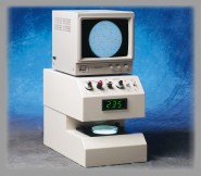
AccuCount™ 1000
The AccuCount™ 1000 is ideal for the AMES Assayusing 35 to 100 mm petri dishes or 6, 12, and 24 format multi-well dishes. The transmitted illumination can be used whenever analyzing objects on a translucent or transparent background. Transmitted darkfield capability assists in analyzing transparent or low contrast objects. The reflected illumination makes it possible to analyze objects on opaque backgrounds, such as blood and chocolate agar.
The AccuCount's outstanding sensitivity and resolution permits counting of a wide range of object types and sizes, including bacterial colonies, cells, and industrial particles.
|
AccuCount™ 2000 automated colony counter
Microscopic Automated Colony Counter
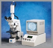
AccuCount™ 2000
The AccuCount™ 2000 automated counter is designed to automatically count microscopic objects in a field displaying totals on a highly visible digital readout. Its outstanding sensitivity and resolution permits counting of a wide range of object types and sizes, including bacterial colonies, cells, grains and industrial particles.
The AccuCount™ 2000 is ideal for scoring Unscheduled DNA Synthesis slides and the Mammalian Cell Mutation Assay. The external video camera can be easily interfaced to a wide number of microscopes.
The AccuCount™ series accessories.
|
PART No.
|
DESCRIPTION
|
|
4-101-0024
|
35 mm MASKING PLATE
|
|
4-101-0025
|
60 mm MASKING PLATE
|
|
|
Masking Plates are used to mask the stacking ring from the counter, eliminating any interference to the counts.
|
|
|
|
|
7-196-0002
|
18 mm LENS
|
|
|
The size of the Lens determines the minimum size object the counter can detect.
|
|
|
|
|
0-110-0003
|
UNIVERSAL TISSUE CULTURE STAGE
|
|
|
The Universal Tissue Culture Stage ensures correct placement and alignment of a tissue culture flask while counting. The Universal Dish Stop ensures correct placement and alignment of petri dishes while counting.
|
System Description - AccuCount™ 1000 and 2000
The systems consists of two units, a main unit and a video monitor. The main unit contains an illumination stage upon which the petri dish or objects to be counted are placed. An integrated CCD video camera scans the objects on the illumination stage and displays a total count of objects. The video monitor displays the objects to be counted and guides the operator in making the initial instrument adjustments. Each object is superimposed with a bright white dot to indicate that it is detected and will be counted. The use of the size control knob allows the operator to select the size of objects to be counted.
System Description - AccuCount™ 2000
The system is functionally identical to the AccuCount™ 1000 except that the main unit does not contain an illumination stage.
Illumination
Objects and their backgrounds vary widely, therefore the operator can select from three methods of illumination to enhance visibility and increase accuracy. 1) Transmitted - for routine objects of high contrast with relatively transparent backgrounds. 2) Reflected - for objects with high contrast with opaque backgrounds. 3) Darkfield - for objects with low contrast with relatively transparent backgrounds.
Object Recognition
The CCD video camera functions as a scanning light detector. An electronic beam moves across the objects in a series of horizontally adjacent lines, generating a continuous video signal. The video signal is compared with a present sensitivity and size threshold established initially by the operator. These thresholds are controlled by the sensitivity and size adjustments. Optimum sensitivity is determined by an operator assisted sensitivity indicator. When the thresholds are exceeded, a pulse is generated by each object. Therefore, the smaller particles and debris which are below the threshold are not detected. After the entire scan is completed, the total thresholds are exceeded, a pulse is generated by each object. Therefore, the smaller particles and debris which are below the threshold are not detected. After the entire scan is completed, the total of signals exceeding the threshold is displayed as the count.
Counting Resolution
The CCD video camera in effect "sees" only one object at a time as it scans across and down the entire illumination area. The minimum object size that can be detected is related to the object diameter, optical magnification and the number of lines in the scanning pattern. The scanning lines are controlled by the CCD video camera and it electronic circuitry. The minimum object size that can be detected also depends upon the contrast between the object and the background. A minimum contrast level of 20% is required to accurately count objects 0.2 mm in diameter. As higher contrast levels are achieved, a minimum object size of 0.1 mm in diameter can be counted.
Closely spaced, clustered or overlapping objects may be counted as multiple or single units, depending on the orientation of the objects to the scan line. In general, such conditions are effectively overcome by employing the compensation factor within the instrument. Another technique is to rotate the subject which contains the objects, recording the average of a few counts at different object orientations. Optional lenses are available to optically magnify the objects by a factor of two, allowing their detection at about one-half of the normal size and spacing requirements.
Aperture
A very simple rapid adjustment at the start of the counting operation is used to establish the size and position of the scanning area. The entire subject may be viewed, but the area that is counted is segmented by the electronic aperture. The aperture is calibrated in square millimeters in the event a statistical comparison of different size areas may be needed. The aperture is viewed on the video monitor and appears as a brighter area superimposed over the subject being counted.
|
APPLICATIONS
|
AccuCount™ 1000
|
AccuCount™ 2000
|
|
MACROSCOPIC COUNTING (> 0.1mm)
|
•
|
|
|
MICROSCOPIC COUNTING (<0.1mm)
|
|
•
|
|
AMES TESTING
|
•
|
|
|
ANTI-BODY PRODUCING CELL ASSAYS
|
•
|
|
|
AUTORADIOGRAPHY GRAIN COUNTING
|
|
•
|
|
BACILLUS BACTERIAL COLONY COUNTING
|
•
|
|
|
BACTERIAL COLONY COUNTING
|
•
|
|
|
BACTERIAL MUTATION ASSAYS
|
•
|
|
|
CLONAGENTIC COLONY COUNTING
|
•
|
|
|
CANDIDIA FUNGI COLONY COUNTING
|
•
|
|
|
COLIFORM COLONY COUNTING
|
•
|
|
|
E. COLI BACTERIAL COLONY COUNTING
|
•
|
|
|
AgNOR's - Ag STAINED NUCLEAR ORGANIZING REGIONS
|
|
•
|
|
MAMMALIAN CELL COUNTING
|
•
|
|
|
MOUSE LYMPHOMA ASSAY
|
•
|
|
|
NUCLEATED CELL COUNTING
|
|
•
|
|
PARTICLE COUNTING
|
•
|
•
|
|
PLAQUE FORMING CELL ASSAYS
|
•
|
|
|
SALMONELLA BACTERIAL COLONY COUNTING
|
•
|
|
|
STAPHYLOCOCCUS BACTERIAL COLONY COUNTING
|
•
|
|
|
SURFACE TESTING
|
•
|
•
|
|
VIRAL COLONY COUNTING
|
•
|
|
|
WATER TESTING
|
•
|
|
|
Article No.
|
Title
|
|
CLC1001
|
Evaluation of an Automated Colony Counter
|
|
|
W. A. GOSS , R. N. MICHAUD, AND M. B. McGRATH
|

|
An automated colony counter was found to readily detect surface and subsurface bacterial colonies of 0.3 mm size or greater with a high degree of precision. On a logarithmic scale, counting efficiency consistently ranged from 89 to 95% of corresponding manual count determinations for plates containing up to 1,000 colonies. In routine application, however, automated plate counts up to approximately 400 colonies were selected as a more practical range for operation. The automated counter was easily interfaced with an automated data acquisition system.
|
|
|

|
|
CLC1002
|
The Use of Adult Rat Liver Cultures in the Detection of the Genotoxicity of Various Polycyclic Aromatic Hydrocarbons
|
|
|
C. TONG, M. F. LASPIA, S. TELANG, AND G. M. WILLIAMS
|
|
|
The hepatocyte primary culture (HPC)-DNA repair test and the adult rat liver epithelial cell (ARL)-hypoxanthine-guanine phosphoribosyl transferase (HGPRT) mutagenesis assay are two in vitro short-term tests that possess intrinsic capability for xenobiotic biotransformation. Both assays detected the genotoxicity of a variety of carcinogenic polycyclic aromatic hydrocarbons. Thus, these two tests, which embody intact cellular metabolism, are useful for the evaluation of this class of carcinogens and provide results that strengthen those obtained in tests dependent upon subcellular metabolism.
|
|
|
|
|
CLC1003
|
Health Effects Of Chemicals: V. Computer-Assisted Genetic Toxicology Testing
|
|
|
LEON S. OTIS, ROSIE Mc CORMICK, AND BETTY STROMNESS
|
|
|
Until recently manual grain counting took hours for each slide and made routine testing with this procedure impractical. With the new automated system, grains are counted using a "colony counter," which can detect the exposed silver grains using a contrast-discriminator. Grain counting, therefore, can be accomplished in the time it takes to focus the microscope on a cell, set an electronic aperture over the desired counting region (which is displayed on a TV monitor), and push a count button on the colony counter. The data are fed directly into a file in the computer, eliminating the need to record raw data manually. This system has reduced the time required to score 50 cells per slide from a few hours to an average of less than 10 minutes. In addition, all subsequent processing of data is accomplished by computer programs with no additional entry of data required.
|
|
|
|
|
CLC1004
|
A Rapid, Semi-Automated Counting Procedure for Enumeration of Antibody-Forming Cells in Gell and Nucleated Cells in Suspension
|
|
|
DAVID H. KATZ, MERYL FAULKNER, LEE R. KATZ, ERIK LINDH, CHARLES C. LEONHARDT, KIMBERLEY HERR AND AMAR S. TUNG
|
|
Since the description by Jerne et al (1) of the hemolysis-in-gel technique for enumerating single antibody-producing cells, this procedure has become one of standard usage in most laboratories engaged in immunological research. Different laboratories use a variety of modifications of the technique, whether in a gel support medium as initially described or the suspension technique described by Cunningham and Szenberg (2). Irrespective of the modification employed, the final analysis involves enumeration of plaques which have developed in the indicator erythrocyte suspension. Typically, this has been done visually by examining either slides or petri dishes with an appropriate light source either with or without the aid of a suitable magnifying lens or stereozoom microscope.
|
|
|
|
|
CLC1005
|
Purification and Properties of a Rat Liver Protein that Specifically Inhibits the Proliferation of Nonmalignant Epithelial Cells from Rat Liver
|
|
|
JAMES B. McMAHON, JAMES G. FARRELLY, AND P. THOMAS IYPE
|
|
In inhibitor of cell proliferation was purified from rat liver by alcohol precipitation, ultrafiltration, and DFAE-cellulose chromatography. The hepatic proliferation inhibitor was shown to be pure by polyacrylamide gel electrophoresis in the presence of sodium dodecyl sulfate, analytical isoelectric focusing, and high-performance liquid chromatography. The hepatic proliferation inhibitor was found to have a'molecular weight of 26,000 and an isoelectric point of 4.65. This protein inhibited the proliferation of nonmalignant rat liver cells in culture, and removal of the protein reversed the inhibition produced by low doses. It exerted no effect on the proliferation of malignant rat liver cells.
|
|
|
|
|
CLC1006
|
Semi-Automated Grain and Cell Counting
|
|
|
PAUL A. BRUNN JR., SHARON S. FORD, STANLEY E. SHACKNEY
|
|
A commercially available bacterial colony counter has been adapted for the counting of radioautographic grains over individual cells in smears, and for counting cells in histologic sections. For the counting of radioautographic grains, the correlation coefficients between counts obtained visually by two observers, and between counts obtained visually and using the instrument were similar (r = .999 and r = .998 respectively). The instrument counts were obtained more rapidly than the visual counts and were associated with less observer fatigue. While performance of the instrument in counting cells in mouse bone marrow sections was less accurate than in counting radioautographic grains, a good estimation of marrow cell number was obtained (r = .968). Data on bone marrow cellularity was obtained far more rapidly than with semi-quantitative methods.
|
|
|
|
|
CLC1007
|
Semi-Automated Autoradiographic Measurement of DNA Repair In Normal and Xeroderma Pigmentosum Cultured Human Fibroblasts
|
|
|
KENNETH H. KRAEMER, JOSEPH K. BUCHANAN, AND SHERMAN F. STINSON
|
|
Assessment of DNA repair in cultured human fibroblasts by autoradiography may be facilitated by using semi-automated grain counting instruments. The instrument determined number of autoradiographic grains per nucleus in cultured human skin fibroblasts was found to be linear in comparison to visual counts up to only 30 grains per nucleus. However, with two different instruments a greater range of linearity (100 to 120 grains per nucleus) was attained by measuring the grain surface area per nucleus. Semi-automated analysis of the grain surface area per nucleus yielded measurements of relative rates of unscheduled DNA synthesis after ultraviolet irradiation in xeroderma pigmentosum and normal human fibroblasts, which were reproducible and rapid.
|
|
|
|
|
CLC1008
|
From Image to Analysis: Object Detection and Measurement
|
|
|
MICHAEL K. BENDER
|
|
Image Analyzers, both manual and automated, allow users to identify objects and to categorize I them using various geometric parameters. Densitometric identification and categorization is also possible with some image analysis equipment. Although image analyzers are not a recent development, many of their applications are new. Technological developments in the last decade have extended the capabilities of image analyzers by increasing speed, accuracy, and flexibility. The instruments have also become significantly more affordable.
|
|
|
|
|
CLC1009
|
Detection, Enumeration, and Sizing of Planktonic Bacteria by Image-Analyzed Epifluorescence Microscopy
|
|
|
MICHAEL E. SIERACKI, PAUL W. JOHNSON, AND JOHN McN. SIEBURTH
|
|
Epifluorescence microscopy is now being widely used to characterize planktonic procaryote populations. The tedium and subjectivity of visual enumeration and sizing have been largely alleviated by our use of an image analysis system consisting of a modified Artek 810 image analyzer and an Olympus BHT-F epifluorescence microscope. This system digitizes the video image of autofluorescing or fluorochrome-stained cells in a microscope field. The digitized image can then be stored, edited, and analyzed for total count or individual cell size and shape parameters. Results can be printed as raw data, statistical summaries, or histograms. By using a stain concentration of 5 mg of 4'6-diamidino -2-phenylindole per ml of sample and the optimal sensitivity level and mode, counts by image analysis of natural bacterial populations from a variety of habitats were found to be statistically equal to standard visual counts. Although the time required to prepare slides, focus, and change fields is the same for visual and image analysis methods, the time and effort required for counting is eliminated since image analysis is instantaneous. The system has been satisfactorily tested at sea. Histograms of cell silhouette areas indicate that rapid and accurate estimates of bacterial biovolume and biomass will be possible with this system.
|
|
|
|
|
CLC1010
|
Automated Radiographic Grain Counting: Correction for Grain Overlap
|
|
|
W.H. SCHUETTE, S.S. CHEN, S.J. OCCHIPINTI, H.S. MUJAGIC, AND S.E. SHACKNEY
|
|
|
An algorithm is described for the calculation of radioautographic cell grain count from measurements of total cell nuclear area and total grain area. This algorithm provide-s a statistical correction for grain overlap that is based on the solution to the occupancy problem in probability theory. This method permits the use of automated grain counting over a wide range of grain counts/cell, and extends the useful dynamic range of radioautographic grain counting to well over 200 grains/cell.
|