产品说明
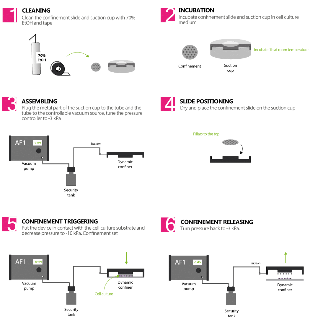
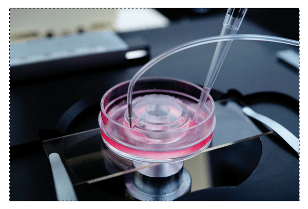
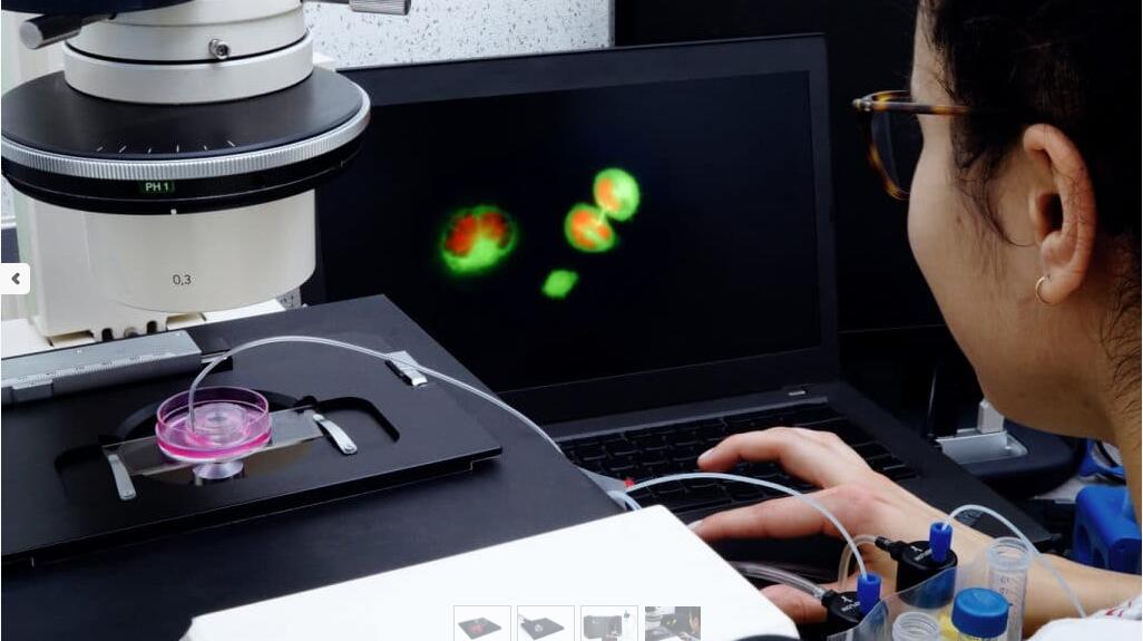
动态细胞限制器是一种将两个平行表面之间的细胞限制在定义的微米高度且具有亚微米精度的装置。两个表面之间的空间由微型 PDMS 柱控制。微柱在载玻片上制造,载玻片连接到 PDMS 活塞(吸盘)。活塞由真空泵控制,因此也控制了限制的高度。
> 控制分娩速度
通过真空泵精雀控制密闭速度
> 可逆限制:限制后取回您的细胞
由于细胞的非破坏性方法,允许进行分子分析
> 与您自己的实验兼容
confiner 是一种小型设备,可直接放置在细胞培养物的顶部
DYNAMIC CELL CONFINER 套件包括:
> 1 x Elveflow 真空压力发生器 + 1 x Elveflow 真空压力控制器
> 12 x Confinement Slides – 10 mm(直径)载玻片/盖玻片,带有 PDMS(支柱)中的微结构,可以进行限制。
可用的限制高度 - 从 1、2、3、... 到 20 ?m(zui多选择 3 个高度来集成您的套件)
> 3 x Dynamic Confiner?(PDMS 吸盘)
> 1 x安quan罐(用于保护真空泵)
> 1 x管和连接器套件
如果您想让幻灯片贴在您的细胞上或不贴在您的细胞上(可选):
> 用于细胞粘附的细胞外基质蛋白(例如纤连蛋白)的等分试样在正确的缓冲溶液中
> 抗粘连分子(聚乙二醇)的等分试样已准备好与载玻片结合
应用
癌症
> 转移细胞的迁移
> 转移中的细胞收缩性
> DNA DSB 修复(机械诱导)
> 基因组不稳定性(细胞分裂)
> 分离的共培养
免疫学
> 免疫细胞的迁移
> 非粘附细胞成像
器官生理学
> 癌细胞的迁移
> 带有刚度控制的细胞分化
> 伤口愈合
> 分离的共培养
> 细胞压缩响应
罕见病
> 细胞核完整性
老化
> 细胞核完整性
> 自噬相关疾病
观察you化
> 非粘附细胞成像
> 细胞器的平面成像
基础研究
> 细胞体积(细胞周期)
> 细胞拉伸反应
文献:
> A flagellate-to-amoeboid switch in the closest living relatives of animals
Brunet. T., et al., Elife, 2021 Jan 15;10:e61037
> The nucleus measures shape changes for cellular proprioception to control dynamic cell behavior
Venturini. V., et al., Science, 2020, 370, eaba2644
> Confinement and Low Adhesion Induce Fast Amoeboid Migration of Slow Mesenchymal Cells
Y.-J. Liu, M. Piel, et al., Cell, 2015 160(4), 659-672
> Actin flows induce a universal coupling between cell speed and cell persistence
P. Maiuri, R. Voituriez, et al., Cell, 2015 161(2), 374–386
> Geometric friction directs cell migration
M. Le Berre, M. Piel, et al., Physical Review Letter 2013 111, 198101
> Mitotic rounding alters cell geometry to ensure efficient spindle assembly
O. M. Lancaster, B. Baum, et al., Developmental Cell, 2013 25(3), 270-283
> Fine Control of Nuclear Confinement Identifies a Threshold Deformation leading to Lamina Rupture and Induction of Specific Genes
M. Le Berre, J. Aubertin, M. Piel, Integrative Biology, 2012 4 (11), 1406-1414
> Exploring the Function of Cell Shape and Size during Mitosis
C. Cadart, H. K. Matthews, et al., Developmental Cell, 2014 29(2), 159-169
> Methods for Two-Dimensional Cell Confinement
M. Le Berre, M. Piel, et al., 2014, Micropatterning in Cell Biology Part C, Methods in cell biology, 121, 213-29
|
