| 产品名称 | Low-Cost Fluorescence Stereo-Microscope Multi-Fluorophore System |
| 产品货号 | |
| 产品价格 | 现货询价,电话:010-67529703 |
| 产品规格 | |
| 产品品牌 | Tritech ResearchInc |
| 产品概述 | Low-Cost Fluorescence Stereo-Microscope Multi-Fluorophore System |
| 产品详情 |
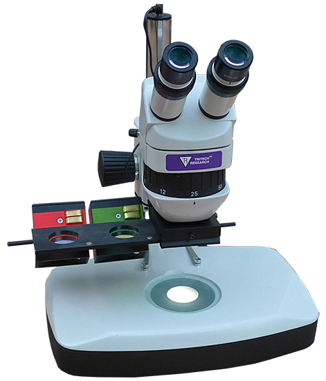 (Click to see larger image) If you got here from a link you saw on Twitter, click here to follow us for more deals and promotions! Because you're a scientist, we've included a lot of details about our new product so that you can understand it and compare it with others, but if you'd like to talk with a person, just pick up the phone and call our tech support staff, 24/7. The SMT1-FL-QC (Pats. pending) is the culmination of our years of research to bring a research-grade, full-featured, low-cost fluorescence dissecting stereomicroscope to the scientific community that is affordable to the education community as well. It allows for rapid switching between different fluorophores like GFP and RFPs in under 1 second and is reconfigurable between multiple fluorophores in just seconds with no tools! With our technology, excitation sources are incredibly bright and background fluorescence is minimized, so dim signals are often visible and many phenotypes can be seen even in a brightly lit room. This is not a mediocre add-on kit with lots of compromises. This is a complete fluorescence microscope system for less than the price of other high quality plain dissecting stereomicroscopes! If you are considering the Zeiss Axio Zoom.V16, the Leica MZ10 F, M205 FA, M165 FC, the Nikon SMZ25, SMZ18, or the Olympus SZX16, MVX10, or SZX10, you really need to take a look at our SMT1-FL-QC to see how much money you can save - not only on purchase price but on maintenance costs while gaining increased equipment life span. Before getting into the great features of our low-cost fluorescence dissecting scope, we want you to know that our better prices do not equate to lower quality they derive from superior engineering and a different company philosophy. When someone buys a microscope from the "Big 4", they are not just paying for the microscope they are paying for tons of advertising, marketing, sales commissions and other hidden costs. At Tritech Research, we forgo all of those expenses and rely on great word-of-mouth recommendations within the scientific community so your precious grant budget or start up funds can be stretched further since they only pay for the microscope itself! Tritech Research isn't a start up; we've been selling dissecting stereomicroscopes for over 25 years, since our inception in 1991. We operate on a lean budget and put money from your purchase back into Research and Development of new products to save even more money for the scientific community while delivering new enabling technologies like this microscope!  How does it work? First, we replaced the expensive mercury arc lamp, power supply, and bulbs that cost about $200 and last only 200 hours with super-bright, energy-effcient LEDs that last about 40,000 hours. We've sourced the brightest LEDs in the world, and designed optics to harvest almost every photon relevant to exciting your fluorescent samples. Our modules employ two excitation beams that intersect in the focal plane of your specimen. This doubles the signal without doubling the background fluorescence* (Pats. Pending). Each compact, interchangeable module uses the finest dichroic filters to refine the excitation and emission wavelengths for bright, clean imaging and a powerful magnetic system for easy, tool-free installation and changing of configuration to suit any single, double, or triple labeled experiment. While playing with the magnets above, we invented a slider system uses magnetic detents (Pats. Pending) for robust, wear-free operation. A single sliding operation precisely positions the excitation and emission filters, and energizes the module. The microscope is available with a few different options for the base/stand and illumination. Fluorescence illumination is always from the top, and we have options that allow for different types of standard white-light illumination of your specimens. For top lighting, select our standard "ergonomic" base and add our dual goose-neck LED illuminator. For transillumination (through the sample), you can use our standard "ergonomic" base with frosted glass plate and built-in fixed LED illumination of ∼30mm in diameter. For an upgraded transillumination option, select our taller, narrower, triangular transillumination base with a large rotatable and translatable mirror. This upgraded base employs a larger frosted mirror and clear glass plate. Moving the diffusion farther from the specimen increases contrast and resolution. The larger mirror can illuminate a sample ∼50mm in diameter, and changing its angle and position allows for optimal contrast to visualize internal structures (for example structures within the C. elegans pharynx and embryos, or within internal organs of Zebrafish, Xenopus, chick and mammalian embryos). Click here to see the triangular base on our non-fluorescence scope with an old-school halogen fiber-optic illuminator. An optional foot pedal allows for the white illumination to be flashed on as needed to orient samples without moving your hands or losing your dark-adapted vision. As this is a Tritech brand product, it is covered by our money-back guarantee and limited warranty. Order it with confidence and try it in your own lab or classroom. If you have concerns about being able to visualize a particular sample, you can even mail us a sample, and we will e-mail you a photo of what we can see with the scope! Here is a video that shows how easy it is to assemble the scope and use it: Here are some images of samples examined with the scope:  The nematode C. elegans expressing the [Punc-25::GFP] transgene (juIs76). unc-25 encodes glutamic acid decarboxylase (GAD). UNC-25 is expressed specifically in GABAergic neurons and localizes to cell bodies, axonal branches, and synaptic regions in the 19 type D motor-neurons, most notably shown here in the 13 ventral nerve cord neurons. 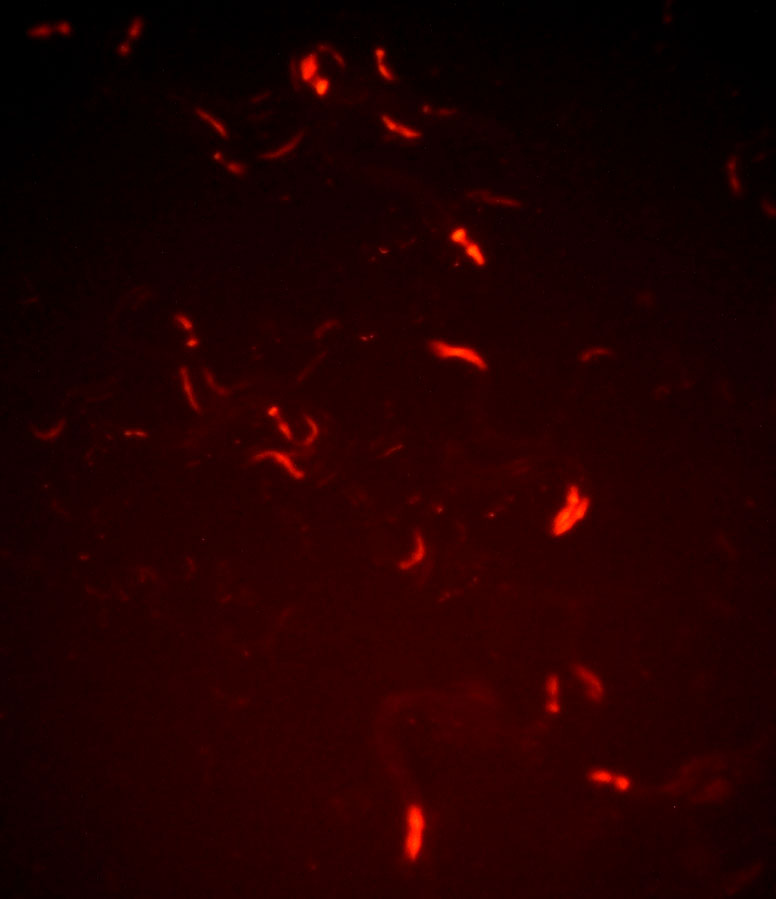 C. elegans expressing a [Pmyo-2::dsRed] transgene. myo-2 encodes a tissue-specific myosin class II heavy chain. MYO-2 is expressed specifically in the muscles of the pharynx, as shown here by the expression of the Red Fluorescent Protein dsRed.  Larvae of transgenic Drosophila melanogaster expressing GFP in their salivary glands. "Tritech Research is the first and only company to offer LED-based complete GFP Dissecting Stereo-microscope Systems, saving customers thousands of dollars. Traditional Mercury arc lamp based fluorescent systems cost up to $35000. They have perfected the LED-based GFP microscopy and completely eliminated the mercury arc lamp and associated optics, bringing the price of the microscope to one third of its previous cost!."FAQ's Q: On the SMT1-FL-QC I noticed that there is a partial occlusion of the field of view in the left eyepiece (like a mild waning gibbous moon). This only is visible at 6x magnification - I can see the whole field clearly at higher magnifications. Why is this? A: The module is the whole unit with the colored emission filter holder plus the dual excitation light source. But technically, the part than may be twisted or jacked up/down is the black box containing the dual excitation light source, relative to the colored emission filter holder. The illuminated spot is purposely smaller than the 6x field of view. At 12x, the edges are a little dimmer, and at 25x + the field should be fully illuminated. The best, brightest images will be seen at 12x and 25x. Hopefully the modules were still aimed perfectly when they arrived. If you're careful when you install and remove them, you won't have to re-aim anything. Sometimes the "slider holder" (the black part under the objective that the slider slides through) can shift in angle if someone pulls down on one side of the slider. In that case both modules will still illuminate the same spot, but it will be off-center.... and then you should fix the slider-holder instead of aligning the modules. Perhpas you are not referring to the portion of the 6x field of view that is illuminated by the converging beams, but instead you are referring to the entire field of view (for example what you see when the sample is illuminated from the bottom. If that is the case, it sounds like the slider is just not exactly stopped in the center of its magnetic detent. Try manually sliding the slider left and right while looking through the scope and see if you can get rid of the gibbous. |
| 产品资料 |