|
|
-
|
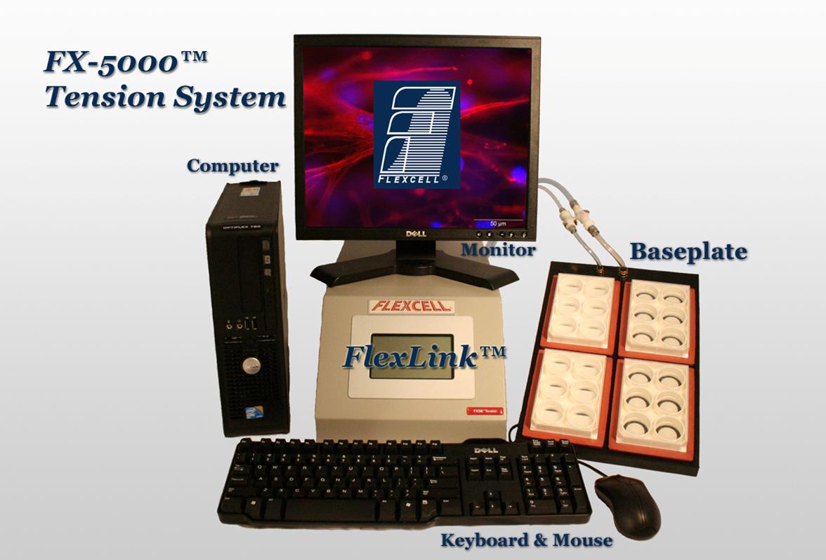
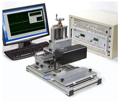
| flexcell® FX-5000™ cell Tension loading culture System |
Features:
A computer-regulated bioreactor that uses vacuum pressure to apply cyclic or static strain to cells cultured on flexible-bottomed culture plates.
1)Apply equibiaxial or uniaxial tension to cells in 2D and 3D culture.
2)Uses regulated vacuum pressure to deform flexiblebottomed culture plates producing up to 33 % substrate elongation.
3)Simulate in vivo tissue strains and frequencies in cells from muscle, lung, heart, blood vessels, skin, tendon, ligament, cartilage, and bone.
4)Contains state-of-the-art digital valve to automatically regulate and maintain pressure to provide the specified strain regimen.
5)Works with BioFlex, TissueTrain, UniFlex series culture plates...
|
|
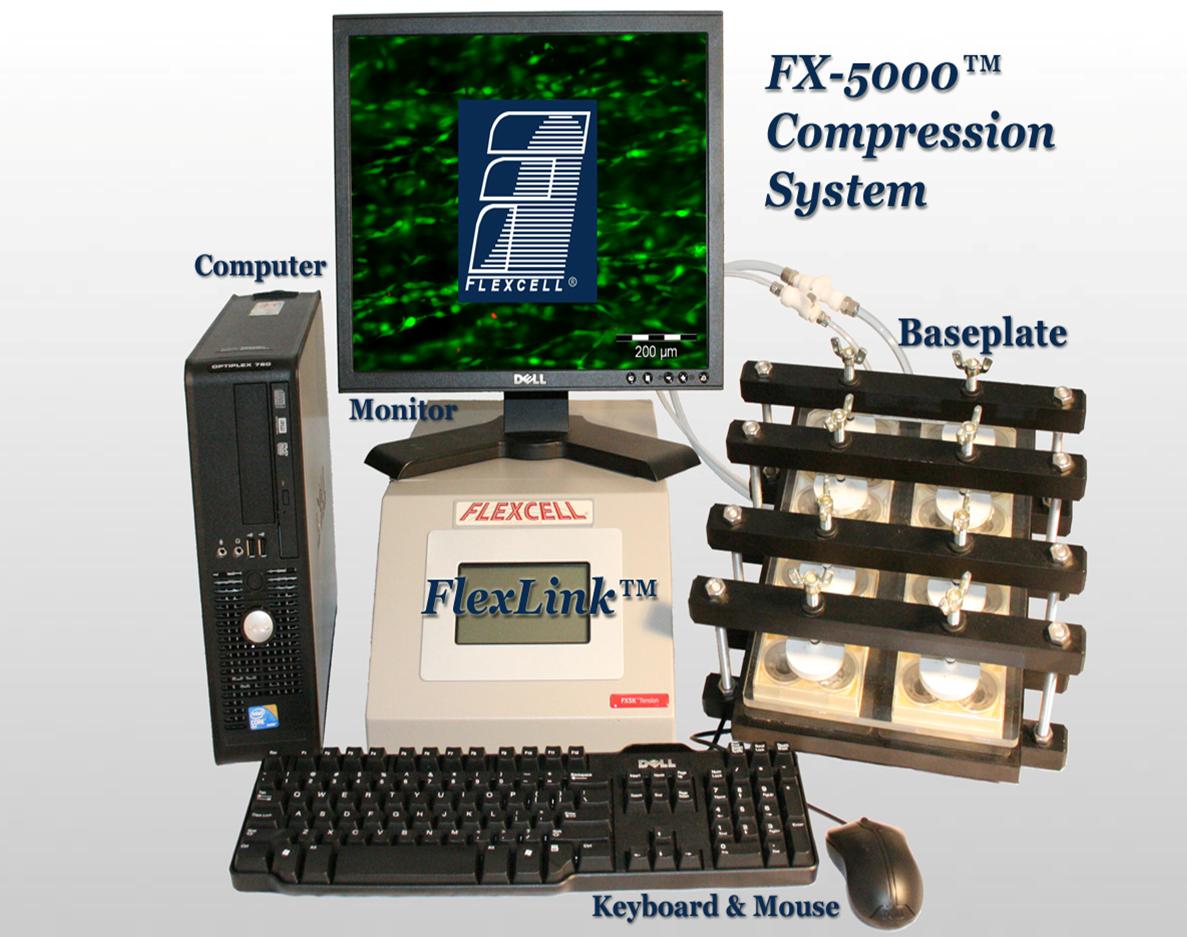
| flexcell® FX-5000™ cell Compression loading culture System |
Features:
A computer-regulated bioreactor that uses positive air pressure to deform tissue samples or 3-D cell cultures in vitro, simulating biological compression conditions.
Uses positive pressure to compress samples between a piston and stationary platen on the BioPress™ culture plate yielding up to 14 lbs of force.
Program multiple frequency, amplitude, and wave changes in one regimen with defined, static or cyclic deformation of cells.
Valving mechanism regulates pressure to provide specified compression regimen..
Compatible with Windows 7.
|
|
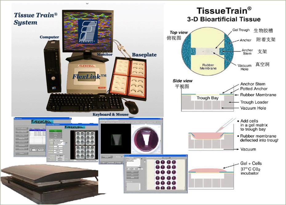
| flexcell® Tissue Train® Culture System |
Features:
A computer-regulated bioreactor that allows users to create 3-dimensional cell-seeded gels and apply uniaxial tensile strains to these gels.
3-D culture in a gel matrix with or without cyclic tension.
Build bioartificial tissue strips of skin, tendon, ligament, etc. up to 35 mm in length.
Observe responses of cells cultured in 3-D matrices with phase contrast, fluorescent, or scanning confocal microscopy.
Compatible with Windows 7.
|

|
| flexcell® Streamer® Shear Stress Device |
Features:
A shear stress device that allows users to apply fluid shear stress to cells in monolayer culture.
Culture your cells on matrix-treated 25 mm x 75 mm x 1 mm Culture Slips®.
Regulate shear stress from 0 to 35 dynes/cm2 using a computer-controlled peristaltic pump.
Works with Osci-Flow® flow controller to regulate pulsatile and oscillatory fluid flow.
|

|
| Permeabilized Myocyte Test System |
Features:
|

|
| 1500A - Small Intact Muscle Test System |
Features:
|
|
| 1310A - Large Rodent,Small Animal Muscle Test System |
Features:
|
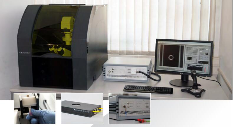
| Optical Stretcher |
|
Features:
The Optical Stretcher is a novel laser tool to measure and analyse biomechanical properties, such as elasticity and relaxation, of single biological cells in suspension.
1)Contact-free cell deformation
Contact-free cell deformation” open=”0″ style=”2″]Suspended cells are deformed by optical forces, leading to absolutely contact-free measurements. This ensures homogeneous cell handling and avoids artefacts due to contact-induced cellular reactions.
2)High-throughput single cell rheology
By integration of a microfluidic system about 300 cells/hour can be measured with ease. This allows for the first time to collect significant statistical data from cell rheology.
3)Timesaving, automated measurements
Cells are automatically delivered to the measurement region and deformed corresponding to the user-defined stretch pattern. While the Optical Stretcher runs the experiment you can focus on interpreting results.
4)included microfluidic system with two pressure controlled channels
5)Ytterbium fiber laser with max. power of 2 W per fiber
6)Installation on inverted phase contrast microscope
7)Housing for laser safety and temperature controlled condition
8)Optional combination with fluorescence microscopy
|
3.Chiaro Nanoindenter
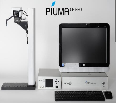
Features:
Compatible with microscopes
The Chiaro Nanoindenter is designed to be compatible with almost any inverted microscope to enable the simultaneous practice of advanced microscopy and micro-mechanical characterization.
Features Chiaro Nanoindenter
The Chiaro Nanoindenter is engineered with flexibility in mind. Due to the almost limitless mounting options and low footprint of our nanoindentation probes, the Chiaro can be combined with many microscopes or other optical set-ups, even simultaneous with other instruments such as electrophysiology equipment, patch clamping instruments, perfusion or heating stage chambers, and many more!
Standard versus dynamic
The standard version of the Piuma and Chiaro Nanoindenter includes all the tools required for performing displacement-controlled experiments, including automatically mapping the micro-mechanical properties of surfaces. The Piuma and Chiaro Nanoindenter are available with a dynamic module, expanding the modes of operation with load-, indentation-, and frequency control.
|
4.Piuma Nanoindenter
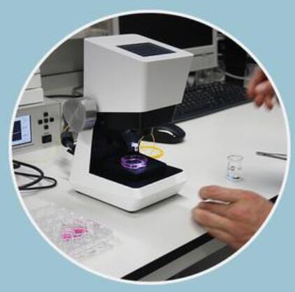
The Piuma Nanoindenter is designed to operate as a stand-alone instrument in any lab setting. Amongst many applications possible, the Piuma can be used to perform comparative studies on biological samples such as cartilage, dermis or cornea, optimize and select between polymer or hydrogel synthesis or crosslinking procedures or define material performance parameters such as surface adhesion, swelling or degradation, also in liquids.
Features:
Easy operation due to the built-in camera (top-view, 5X) and the coarse manual Z-stage. Almost any sample size or shape can be used thanks to the large sample stage. Optionally a sample stage heater is available (from room temperature up to 60oC), a back light illumination module, an inverted camera module for small or transparent samples and an acrylic housing for use in more noisy environments.
|
-
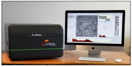
| BioRam®System— Live cells Raman spectroscopy system |
|
Features:
Photonic fingerprinting® for gentle cell analysis,Raman Trapping Microscopy for stress- and label-free cell identification and characterization
1)Identifying cells from their biophotonic profile
Raman spectroscopy is based on the Raman effect: when a molecule is exposed to laser light, a small fraction is scattered with a shift in frequency compared to the incident light. This shift is highly specific for each molecule – as unique as a fingerprint.
Raman spectroscopy has long been used successfully as a contact-free technique for material characterization. More recently, scientists have demonstrated its applicability to the field of biology and medicine: several research teams have independently shown that the combined Raman spectra of all biopolymers within a cell form distinct clusters according to cell type and state of differentiation and activity
2)Mastering the challenges of biological samples
In close collaboration with scientists at the Fraunhofer Institute for Interfacial Engineering and Biotechnology, CellTool have overcome the particular technical challenges that have so far hampered routine use of Raman spectroscopy in biomedical research and development. Biological samples often emit fluorescence which interferes with their weak Raman signals. Furthermore, they require a tolerable wavelength and appropriate physiological conditions during measurement to preserve cell viability.
3)Raman spectroscopy for living cells
BioRam, CellTool’s most advanced analytical platform, is available for customers.
4)The measurement process: Just get going
Samples can be used without special preparation: as living tissue, in aqueous solution on Raman-compatible slides or directly in culture medium in Raman-compatible petri dishes or fluidic chips. With a 3D-mouse, you can home in on the cells to be analysed and start the automatic generation of Raman spectra with a mouse click. Cells are then automatically identified according to their spectra by database comparison. Measurement coordinates, live images, raw data and all the relevant parameters are saved as well, enabling further analysis or the application of alternative algorithms at any time.
|
| Cellsorter®Automated single cell sorting and deposition |

|
Features:
How to get the one you want? Single cells are in your hands——sort live cells right from the petri dish
1)High throughput single cell sorting directly from the Petri dish
2)One single cell arrives to each PCR tube
3)10 PCR strips containing 80 tubes can be filled in a cycle
4)Glass cover slip for testing single cell deposition in situ
5)Drop volume less than 1 ul
6)15-20 seconds per cell
7)Isolates a subpopulation of live adherent cells expressing fluorescent or luminescent markers
8)Both unlabeled and fluorescent cells are recognized by computer vision
9)Viable cells after sorting
10)Sorts cells labeled by fluorescent molecular probes highlighting specific cellular activities
11)Collects single cells for further cultivation, cloning, RNA or protein preparation
12)Sorts fixed cells after immunocytochemical preparation
13)Any adherent cell type can be sorted
14)Cell culture needs minimal preparation before sorting
15)Average sorting process takes only a few minutes
16)Multichannel detection using the fluorescent filter setup of the microscope
17)When collecting multiple cells sorting speed is 1 cell/second. Number of cells picked up in a single run: 1-1000.
|
| calScreener™multi-channel calorimetric system for cellular |
|
Features:
Monitor cell metabolism and cellular bioenergetics in real time with the label-free calScreener™ from SymCe
1)Monitor cell metabolism and cellular bioenergetics in real time
calScreener™ is the first multi-channel calorimetric system specifically developed for cell based assays. Measurement of heat flow in living cells is a well established and proven technology to monitor changes in metabolic processes. calScreener™ provides the researcher with a label-free detection tool with maximal versatility in assay type.
The real time continuous measurement provides important kinetic data for interpretation of cellular mechanisms and responses to treatment.
calScreener™ is suited to drug development and metabolic research providing a true phenotype response where the total status of the cell metabolism is monitored.
calScreener™ is further suitable for toxicology tests, process development and environmental monitoring.
2)Mastering the challenges of biological samples
3)First Calorimeter designed for cell biological work
4)Label-free assay
5)Independent of cell morphology
6)Measures heat produced from cell metabolism
7)True PHENOTYPE response measured
8)Real-Time continuous measurement
9)Prior knowledge of pathways and specific target function not needed
10)Multiple compounds can be tested simultaneously for synergetic effects without knowledge of pathway interactions
11)Suitable for a wide range of applications
|
-

|
| 3D Vascular Bioreactor |
Features:
The DynaGen® Series bioreactors provide mechanical stimulation to three dimensional tissue engineered constructs to create physiological conditions in-vitro. The system imparts pulsatile flow and pressure to a vascular conduit. Applications include the investigation of cell function and differentiation, pharmaceutical benchtop testing, and the seeding and growth of engineered tissues and medical devices
|

|
| HeartValve Bioreactor |
Features:
The DynaGen® Series bioreactors provide mechanical stimulation to three dimensional tissue engineered constructs to create physiological conditions in-vitro. The system imparts pulsatile flow and pressure to a valve or valve-like structure. Applications include the investigation of cell function and differentiation, pharmaceutical benchtop testing, and the seeding and growth of engineered tissues and medical devices.
|

|
|
Ligament and Tendon Bioreactor |
Features:
The DynaGen® Series bioreactors provide mechanical stimulation to three dimensional tissue engineered constructs to create physiological conditions in-vitro. The system imparts axial stress or strain to a ligament or tendon construct. The stress/strain profiles can be defined by the user and can be a simple harmonic (sinusoidal) or a physiological waveform. Applications include the investigation of cell function and differentiation, pharmaceutical benchtop testing, and the seeding and growth of engineered tissues and medical devices. Researchers are currently utilizing these systems in a wide range of research areas including:1)ACL regenerative medicine.2)Collagen gel stimulation.3)Small diameter tendon stimulation
Hand tendon and ligament regenerative medicine
|
|
| OsteoGen Bioreactor |
Features:
's OsteoGen bioreactors impart perfusion through cell seeded cylindrical scaffolds. Applications include investigating cell function, modulating the growth and development of engineered tissues, or acting as a test bed for drug and regenerative medicine technologies. Researchers are currently utilizing these systems in a wide range of research areas including:1)Bone stem cell phenotype research2)Mineral deposition of marrowstromal cells
|

|
| Cartilage Bioreactor |
Features:
The DynaGen® Series bioreactors provide mechanical stimulation to three dimensional tissue engineered constructs to create physiological conditions in-vitro. The system imparts compressive or hydrostatic stress to a cartilage construct. The stress/strain profiles can be defined by the user and can be a simple harmonic (sinusoidal) or a physiological waveform. Applications include the investigation of cell function and differentiation, pharmaceutical benchtop testing, and the seeding and growth of engineered tissues and medical devices. Researchers are currently utilizing these systems in a wide range of research areas including:1)Stem cell stimulation. 2)Meniscus regenerative medicine.3)Chondrocyte stimulation
|

|
| 3D skin Bioreactor |
Features:
The bioreactors impart static and oscillatory stimulation to a "skin like" sample in a unique air-to-nutrient media interface. Applications include investigating cell function and stimulating the growth and development of engineered tissues. The device can also be used as a "test bed" for pharmaceutical and regenerative therapies. The can apply either stress or strain using operator defined wave forms
|
-

|
| E-FIBER ELECTROSPINNING SYSTEM-STARTER KIT |
Features:
E-Fiber electrospinning is a plug & Play setup for users who deal with electrospinning techniques for the first time or for expert users who intend to develop new materials and require a new and easy setup for their research.
The Starter Kit, however simple and easy to use, still has all the requirements and strong points of the high-end versions, such as the Advanced and the XL models.
|

|
| E-FIBER ELECTROSPINNING SYSTEM-DESKTOP BASIC VERSION |
Features:
E-fiber electrospinning advanced version is optimal for research centers, companies and universities whose needs are to produce nanomaterials intended both for commercial and research purposes.
E-fiber is a fully-customisable electrospinning platform consisting of injection stages and application-specific collectors (static or movable), for the laboratory-scale manufacturing of polymeric-nanofibrous matrices. This device creates a powerful and highly versatile technique for the creation of various fibrous architectures with controlled fibre diameter and orientation. Its technology minimises influence on the electrospinning process. E-fiber offers various technological solutions to accurately control each process guaranteeing batch-to-batch reproducibility and precise control of fibre parameters such as diameter, orientation and texture. The equipment is user-friendly and safe thanks to the automatic safety devices that protect the operator.
|

|
| E-FIBER ELECTROSPINNING SYSTEM-XL VERSION |
Features:
The DynaGen® Series bioreactors provide mechanical stimulation to three dimensional tissue engineered constructs to create physiological conditions in-vitro. The system imparts pulsatile flow and pressure to a valve or valve-like structure. Applications include the investigation of cell function and differentiation, pharmaceutical benchtop testing, and the seeding and growth of engineered tissues and medical devices.
|
-

|
| Single Molecule Biophysics Magnetic Trap—Stretching and twisting single molecules |
Features:
PicoTwist is involved in nanomanipulation and single-molecule experiments in biology. Historically, single molecule experiments were among the first nanotechnologies, appearing in the 1990s to characterize biopolymers and proteins with unprecedented accuracy. The idea is that instead of analyzing the behavior of a great number of molecules as usually done in a test tube, you now analyze the behavior of a single molecule. Thus instead of measuring average behavior you have direct access to the biopolymer’s activity in real time.
|
|
| Turnkey TIRF Microscopy complex |
Features:
Turnkey TIRF Microscopy complex is compatible with Nikon, Olympus, Zeiss, and Leica
1)inverted microscopes. The complex includes:
2)Prism-based p-TIRFM flow system, and/or
3)Lightguide-based lg-TIRFM flow system, and/or
4)Objective-based o-TIRFM flow system
5)Multi-color computer-controlled illuminator
6)Low light EMCCD camera Andor iXon or similar
7)Optional digital fluidics SmartFlow
8)Optional temperature control system TC-40 – 25-40oC
9)Optional filter-wheel EW-6 at emission channel
10)Optional electrochemical control unit EC-1070
TIRF Labs supplies turnkey TIRFM complex with TIRF Studio software package, which controls
MCI-7000 illuminator, Andor EMCCD cameras, digital fluidics SmartFlow, temperature control
TC-40, filter-wheel EW-6, and electrochemical unit EC-1070. Microscope is not included. TIRF
Labs will collaborate with microscope supplier of your choice to provide seamless integration of
TIRFM complex with the microscope.
applications:
Cell biology studies
Molecular diagnostics
br>Analysis of biomolecular interactions
Monitoring real-time kinetics of biomolecular interactions
Real-time TIRF microarrays
Combined DNA, RNA, protein, and metabolite arrays
Studies of protein-protein and protein-DNA interactions
Studies of surface supported membranes
Lipid rafts studies
Nanoengineering
Drug screening
Lead optimization
Bioassay development, and more ...
|
|